
Neurofibromas are a type of benign tumor developed from the myelin sheath of the peripheral nervous system and frequently seen in neurofibromatosis type 1 (NF1). Neurofibroma of the head and neck regions is not uncommon.1 There were only a few cases of isolated external ear neurofibroma without neurofibromatosis (NF) reported, as they rarely affect the external ear.1 It is believed that neurofibromatosis is autosomal dominant; the type 1 neurofibromatosis is due to disruptive mutation of the NF1 gene, which codes for protein neurofibromin.
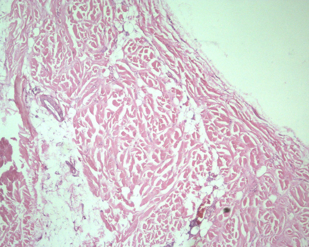
Case reportA 38-year-old Malay male with no comorbidity came to our otorhinolaryngology, head and neck clinic with the complaint of a right postauricular swelling for the past two years. He was more concerned about the cosmetic disfiguring. The swelling was insidious at the onset and progressively increasing in size. It did not cause any pain. There was no history of bleeding from the swelling and no history of any such lesions in the body. No significant family history. Examination of the swelling showed presence of a single 4cm×5cm mass, firm, with regular surface over the post auricular region. Subsequently, we performed a surgical removal of the mass for this patient via a postauricular approach under general anesthesia. The mass was partially adherent to the skin. After removing the excess skin which was adherent to it, the entire mass was completely excised. Histopathological report (Fig. 1) revealed that the lesion was composed of spindle-shaped cells with fusiform nuclei arranged in fascicles. No cellular atypia or necrosis seen. These histological features are suggestive of neurofibroma. Patient has an uneventful recovery, and a good cosmetic result was achieved. There is no recurrence during follow up for the past year.
Discussion
Neurofibromas are a benign neoplasm, derived from the myelin sheath of the peripheral nerve. They may occur in the presence of neurofibromatosis, a hereditary condition.2
There are two types of neurofibromatosis: type 1 (NF1) and type 2 (NF2).
NF1 is an autosomal dominant type, which commonly presents with peripheral nerve sheath tumors called the neurofibromas.3 NF1 is caused by a change in a gene on the chromosome 17. NF2 usually grows into the spinal cord or brain and is caused by a change in chromosome 22. Bilateral vestibular schwannomas of the acoustic nerve and multiple meningiomas are characteristic, rarely with cutaneous manifestations.3,4
There are two types of neurofibromas: dermal and plexiform. Dermal neurofibroma is also called discrete or cutaneous neurofibroma. They usually develop during adolescence and adulthood. They tend to involve the terminal nerves and may be numerous, but there is no apparent risk of malignant transformation. However, despite their benign nature, they may cause significant cosmetic problems and occasionally require removal.
Conversely, plexiform neurofibromas are usually congenital. They developed in childhood and are often extending deeply along the nerves and may involve all levels of skin, fascia, muscle, bone and even viscera. They can become very large and may cause functional impairment, with risk of malignant transformation in 6% in these tumors.4,5
Change in the genetic material that causes NF1 or NF2 can be inherited from a parent, thus referred to as autosomal dominant, or may occur due to spontaneous mutation.1 In our case report, there was no family history. Solitary lesions are not usually associated with the presence of systemic manifestations, unlike multiple lesions, which are commonly seen in patients with NF or von Recklinghausen's disease.4
In our case report, the patient did not have any other features of NF1. Most tumors caused by NF require no treatment; however, when these tumors cause pain, growing rapidly, disfiguring or impairing function, they may need treatment.
Ghosh S.K et al. (2008) reported a case of neurofibroma of the external ear where he removed the entire lesion along with excess skin, and a good cosmetic result was achieved.1
Shaida AM et al. (2007) also reported a case of neurofibroma of the pinna in which surgical excision provided a satisfactory outcome and resulted in an excellent functional and cosmetic outcome.5
In our case, swelling over the postauricular region causing cosmetic deformity was the chief complaint, and we were able to obtain a satisfactory result with no recurrence on follow up for a year.
Final commentsNeurofibromas are commonly seen in a patient with a history of neurofibromatosis. In rare cases of solitary neurofibroma, depending on their size and site of lesion, complete excision is possible with any significant functional and cosmetic deformity. Here, we reported a rare case of neurofibroma, a nerve sheath tumor which presented to us with a swelling behind the ear, which caused cosmetical disfiguring. Complete excision via the postauricular approach gives a satisfactory result with no recurrence, as was achieved in our case.
Conflicts of interestThe authors declare no conflicts of interest.
Please cite this article as: Shi Nee T, Ami M, Min Han K, Sabir Husin Athar PP. Postauricular neurofibroma – a rare occurrence. Braz J Otorhinolaryngol. 2017;83:600–1.
Peer Review under the responsibility of Associação Brasileira de Otorrinolaringologia e Cirurgia Cérvico-Facial.
gology is pleased to honor the reviewers


