
Spontaneous temporomandibular joint (TMJ) herniation into the external auditory canal (EAC) resulting from the foramen of Huschke is a very rare condition, observed in only 0.4% of the population.1 The herniation may mimic the presentations in the osseous wall of the EAC eroded by cholesteatoma, trauma, and other neoplastic or inflammatory lesions.2
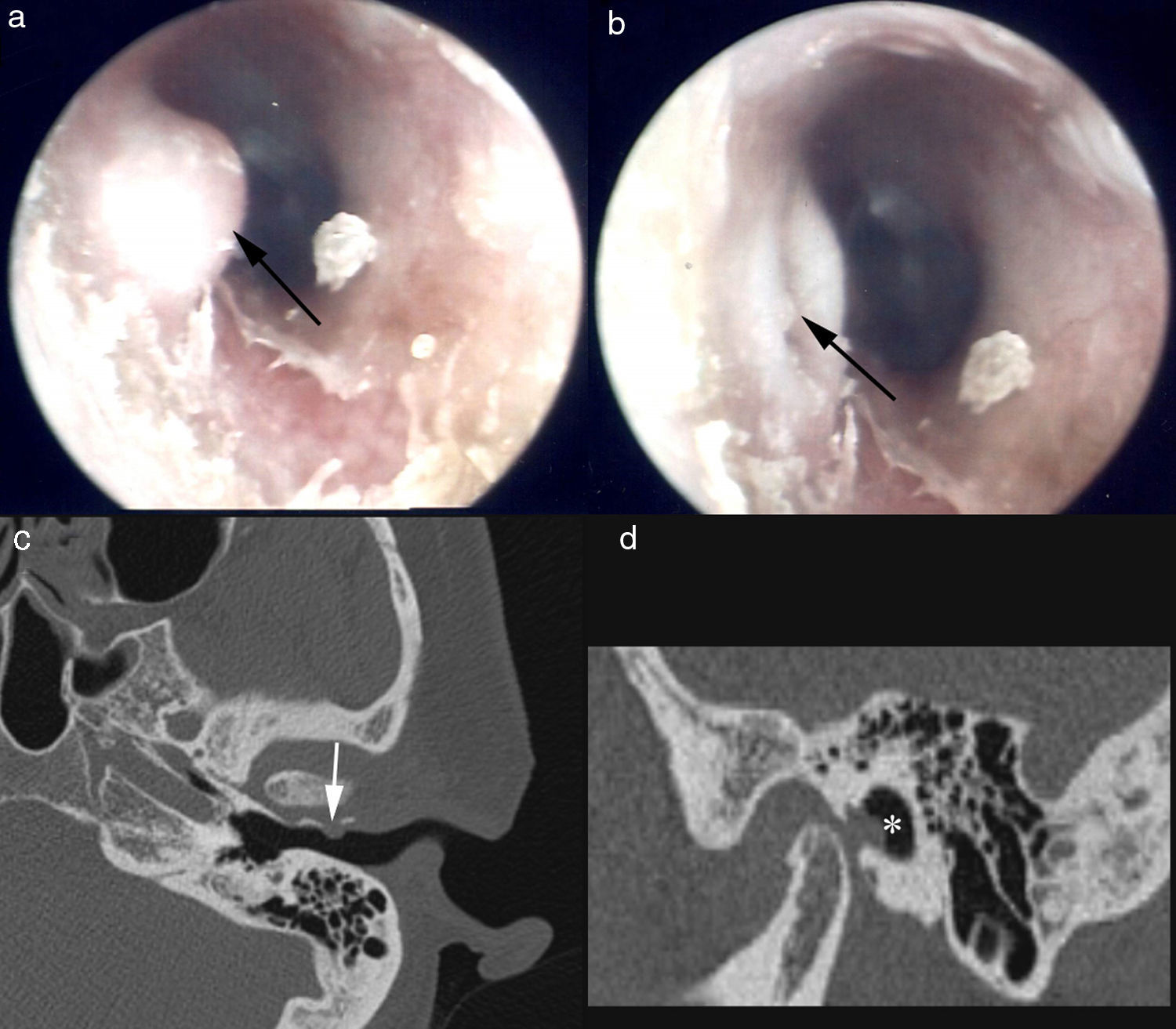
Case reportA 57-year-old woman complained of one-year history of aural fullness and otalgia in her left ear, especially during mastication. She reported no tinnitus, otorrhea, or hearing loss. The patient had no history of ear infection, trauma or surgery. At the otoendoscopic examination, the tympanic membrane was normal but there was protrusion of the soft tissue, originating from the anterior wall of the left EAC. The protrusion appeared as a dome-shaped mass when the mouth was closed (Fig. 1a). When the mouth was open, the mass was retracted anteriorly, leaving an invagination within the meatus (Fig. 1b). A high-resolution computed tomography (HRCT) scan of temporal bone with closed mouth demonstrated a 3mm defect in the anterior wall of the left EAC (Fig. 1c), with a soft-tissue mass extending from the TMJ into the EAC (Fig. 1d). Since it is a benign lesion, and furthermore, the patient did not suffer much inconvenience from the aural fullness and otalgia, we offered a conservative treatment to the patient, and biopsy was avoided. When the patient returned for follow-up one year later, the otalgia had disappeared, while the aural fullness was almost the same as before.
, and retracted when the mouth was open (b). The axial (c) and sagittal (d) high-resolution computed tomography showed a bone defect of the anterior wall of the external auditory canal (arrow) with herniation of soft tissue material (asterisk).")
Otoendoscopic findings showing the protrusion as a dome-shaped mass when the mouth was closed (a), and retracted when the mouth was open (b). The axial (c) and sagittal (d) high-resolution computed tomography showed a bone defect of the anterior wall of the external auditory canal (arrow) with herniation of soft tissue material (asterisk).
In a worldwide literature search, only 25 cases with spontaneous TMJ herniation had been previously described. Their ages ranged from 15 to 87 years, with an average of 56.6 years, and 20 cases (80%) were 50 years old or older. One hypothesis states that the initial dehiscence in the foramen of Huschke would be too small to result in herniation of TMJ or any other soft tissue contents into the EAC. Over time, years of mastication could have softened the intervening tissue or enlarged the foramen with age. This may be the explanation of the age distribution of the reported cases.3 The symptoms in patients with TMJ herniation were nonspecific, but the most common were otalgia (36%), clicking tinnitus (36%,) and otorrhea (32%), followed by hearing loss (20%) and aural fullness (10%); 8% were asymptomatic.
Diagnosis can be established based on otoscopic findings and imaging studies. The herniation is characteristic, because it is more prominent when the mouth is closed and retracts when the mouth is open. Evaluation of mass in the EAC during masticatory movements may be helpful.4 The management of TMJ should be based on the symptoms experienced by the patients as well as on the patient's willingness and suitability for surgery.5 Usually, if patients with TMJ herniations present with trivial symptoms or are asymptomatic, surgery is not considered. However, surgical closure is possible in patients with significant symptoms.6 Of the 25 published cases, 15 underwent surgery: seven (46.67%) cases with clicking tinnitus, six (40%) cases with otalgia, and five (33.33%) cases with otorrhea. For our case, the patient presented with trivial symptoms that which did not bother her too much, and tumor in the EAC was excluded. Conservative management was proposed.
ConclusionWhen assessing patients with mass in the EAC, it is important to consider the possibility of herniation. For most patients with trivial symptoms, a conservative strategy can be chosen.
FundingThis study was supported by the project on Advanced and Frontier Techniques for Shanghai Municipal Hospital (SHDC12010119).
Conflicts of interestThe authors declare no conflicts of interest.
Please cite this article as: Li W, Dai C. Spontaneous temporomandibular joint herniation into the external auditory canal. Braz J Otorhinolaryngol. 2015;81:339–41.


