
Myelomas are plasma cell dyscrasias characterized by lymphoid neoplastic proliferation of B cells, and the multiple myeloma (MM) is the most important symptomatic monoclonal gammopathy. It is characterized by numerous abnormal plasma cells permeating the bone marrow, and overproduction of monoclonal light-chain or heavy-chain immunoglobulins, that are identifiable in serum or urine.1 Because of its tendency to widespread manifestations in multiple organs, this disease interests many medical specialists, including oral and maxillodental surgeons.2 MM manifestations in the head and neck are common, but usually occur on the late stages of the disease; mandibular involvement as the initial presenting sign of the disease is extremely rare.3,4 The aim of this article is to analyze this disorder, based on the presentation of a 67-year-old woman with a painful mass in the mandible that prompted a MM diagnosis.
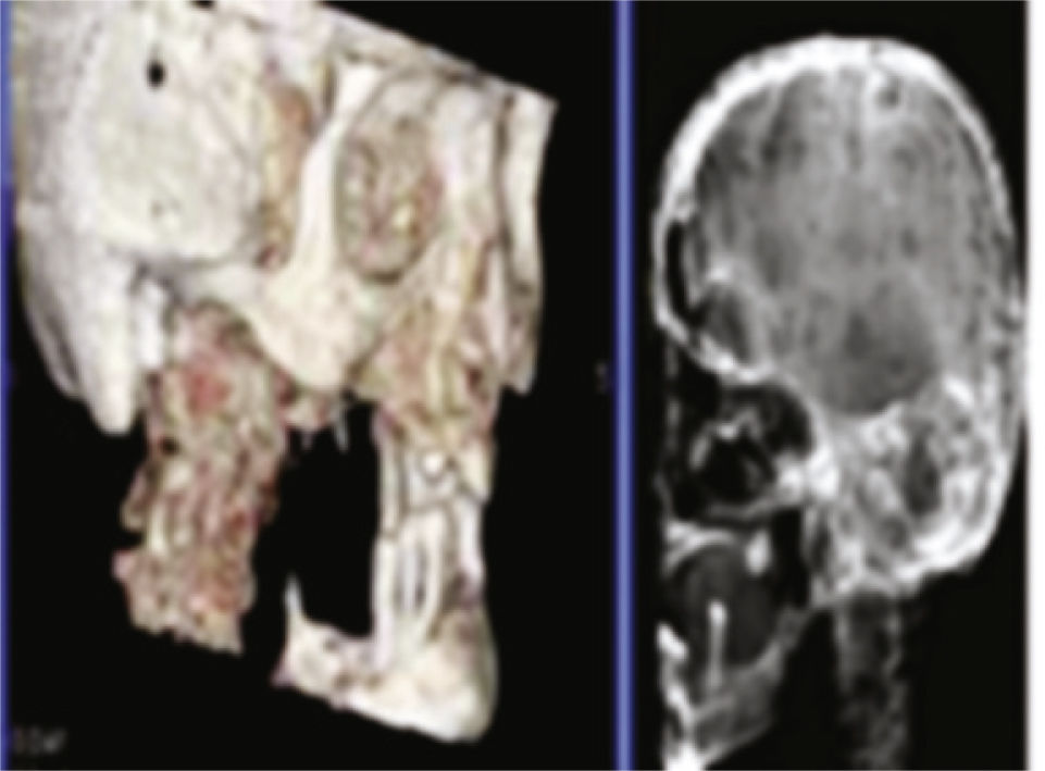
2Case presentationA 67-year-old woman presented a painful and progressively enlarging mass on the right posterior area of lower jaw, which had started approximately one month previously. She experienced dull and intermittent pain on the right side of lower jaw and difficulty in eating. Physical examination showed swelling on the same side of the mandible. Her medical history indicated that she experienced fatigue for the last three months. Intraoral examination revealed a localized soft-tissue mass with central ulceration in right mandibular posterior region. Panoramic radiography showed a large radiolucent lesion of the right mandibular ramus together with multiple smaller “punched-out” lesions. Results of the contrast CT scan confirmed the existence of a large osteolytic lesion in the right mandibular ramus (Fig. 1A). Histopathological examination following incisional biopsy of the soft-tissue mass revealed sheets of plasma cells showing nuclear pleomorphism and lymphocytes, suggestive of plasmacytoma. A whole-body bone scan and a systemic skeletal radiographic survey revealed multiple “punched-out” osteolytic lesions involving the skull (Fig. 1B), clavicles and pelvis. Urinalysis results were positive for Bence Jones proteins. Multiple myeloma was confirmed, and the patient was admitted to receive chemotherapy with proteasome-inhibitor bortezomib in combination with melphalan and prednisone, and to undergo local radiotherapy for the mandibular mass.
3Discussion
Oral lesions are seen with some frequency (30%) in patients with MM; however, when oral lesions are noted, the disease is usually in an advanced stage. Oral manifestations include jaw pain, tooth pain, paresthesia, swelling, soft tissue mass, mobility of teeth, migration of teeth, hemorrhage, and pathologic fracture.2 MM have a high potential to produce local and systemic perturbation of hemostasis and distinct radiographic bone alterations. Undiagnosed MM can impose a formidable emergency condition in dental practice.5 Thorough radiographic examination is critically important because it can be pathognomonic and may be the first sign of the disease.2 The presence of multiple punched-out lesions on the jaw, and the skull bone is also observed in diseases such as Langerhans’ cell histiocytosis, and metastatic malignant lesions.5 MM affecting the jaw bones has a characteristic distribution, more frequently found in the mandible, especially in the posterior region, where hematopoietic activity is greater.4
4Final remarksAwareness of the maxillofacial manifestations of MM is important for early detection of the disease, which may otherwise lead to delays and even errors in diagnosis and treatment. Being a rare disease, MM should not be kept at the forefront of the differential diagnosis of jaw lesions. Rather, we hope the present report will serve to remind us that oral lesions may be the presenting sign of this rare pathology.
5Conflicts of interestThe authors declare no conflicts of interest.
Please cite this article as: Pushpanshu K, Punyani S, Kaushik R. Mandibular mass as the primary manifestation of multiple myeloma. Braz J Otorhinolaryngol. 2014;80:266-7.


